By Sarah Dean,
MSc student at University of Guelph
Marbling can be a challenging trait to achieve, as the degree to which marbling is formed is affected by genetic, sexual, nutritional, and management factors. Generally, most efforts to increase marbling are made during the finishing stage, when high concentrate diets during the finishing stage contribute to marbling. However, other fat depots such as subcutaneous and visceral fat will also increase. These fat depots are often considered ‘waste fat’ whereas intramuscular fat, or marbling, is considered ‘taste fat’. Therefore, the goal is to increase intramuscular fat without increasing subcutaneous and visceral fat. Intramuscular fat is developed during the late stage of pregnancy (third trimester) until 250 days of age (roughly weaning age). This time period is known as the marbling window (Figure 1).
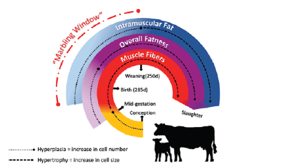
Figure 1: The “Marbling window.” This diagram shows the period of adipose tissue and muscle development in beef cattle from conception to slaughter. Source: Adapted from Cosa et al (2021).
During the marbling window undifferentiated progenitor cells are committing to either myogenic lines or adipogenic lines – in other words, either to become muscle cells or fat cells. In order to increase marbling, we are utilizing vitamin A to increase the commitment to adipogenic lines, or fat cells. When vitamin A is ingested, it becomes retinoic acid. At early stages of development, retinoic acid can regulate the expression of specific genes associated with fat cell development by upregulating the commitment of progenitor cells to become preadipocytes, which likely contribute to marbling deposition later in life. It is essential to increase the intramuscular adipose cells during this time, as after the marbling window, there is no increase in the number of intramuscular adipose cells, they will only increase in size.
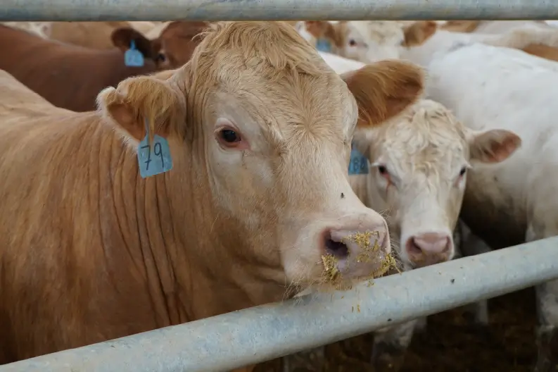
Past research has shown that vitamin A injected into calves at birth had a positive association with marbling. My research is taking this a step further by exploring the supplementation of vitamin A during the third trimester of pregnancy to determine the effect on marbling in the offspring. Thirty angus cross cows pregnant with male calves were randomly assigned to two experimental treatments from 180 gestation until calving: a control group (CON, n=15) receiving 56.76 KIU of vitamin A per day, and a vitamin A supplemented group (VITA, n=15) receiving 227.12 KIU vitamin A per day. Vitamin A was directly incorporated into a pre-mix that was then added to the feed via a TMR mixer. After calving, all cow-calf pairs were raised as a single herd; there were no differences in raising either group and all calves were raised under the same conditions through to slaughter. Within 10 days of calving, skeletal muscle biopsies were collected from the calves to be later analyzed for gene expression and protein abundance of target genes/proteins relating to adipogenesis. Steers were ultrasounded several times throughout their life to monitor the development of intramuscular fat and subcutaneous fat. Steers were slaughtered at 16 months of age when various carcass measurements were collected and evaluated.
Various genes and proteins were evaluated for their expression and abundance to see the impact of higher levels of vitamin A on pathways to create fat cells. There was increased expression of a receptor for retinoic acid (RARβ); indicating that there was upregulation of retinoic acid receptors due to increased retinoic acid being present from feeding more vitamin A. Also, when looking at the genes associated with adipocyte (fat cell) development it was found that there was an increased expression of genes that are likely to lead to an increased number of fat cells being produced (PPARγ and DlK1). DLK1 is associated with the formation of preadipocytes and PPARγ is associated with the commitment of preadipocytes to become mature adipocytes (Figure 2). These results suggest that the presence of more vitamin A, produces more cells that are committed to making intramuscular fat.
Frequent ultrasound imaging showed increased intramuscular fat deposition in vitamin A supplemented calves throughout all evaluated time periods, while there were no changes observed within subcutaneous fat thickness between groups. Hot carcass weight, dressing percentage, carcass size, and internal fat was not affected by treatment. Carcass grades are yet to be analyzed.
These results suggest that vitamin A supplementation during late gestation enhances intramuscular adipogenesis in offspring, meaning the offspring have the potential to produce more marbling and more of a quality meat product. However, the underlying mechanisms of the direct effects of vitamin A on intramuscular adipocyte population requires further investigation, which we will be conducting in the near future.
This article was written for the Spring 2024 Central & Atlantic Beef Grist. To read the whole Beef Grist, click the button below.